Product information "Foot - Superficial and deep dissection of distal leg and foot"
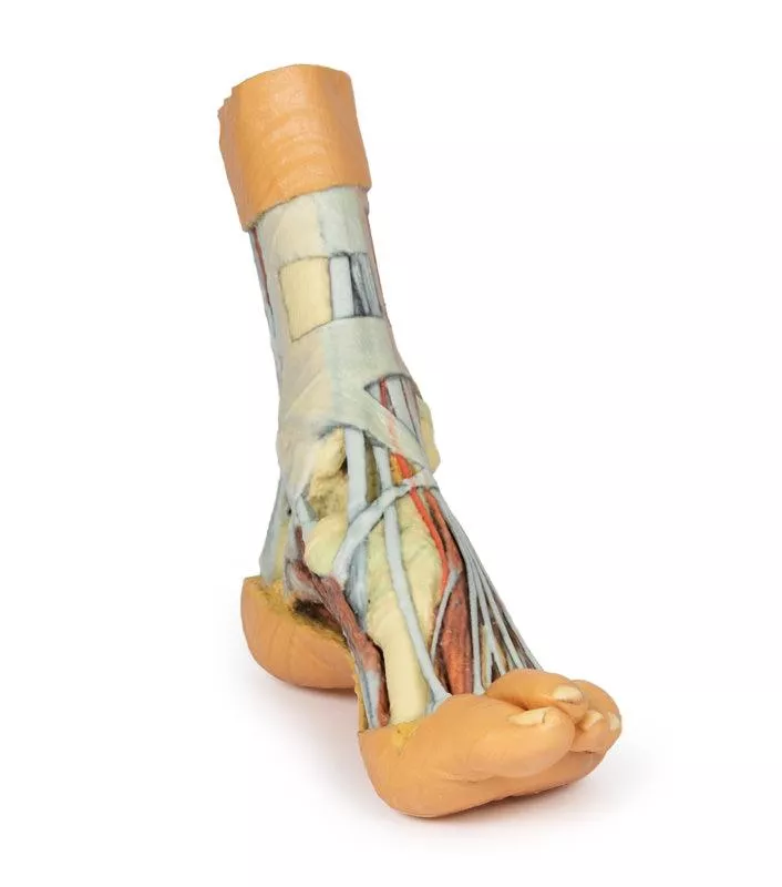
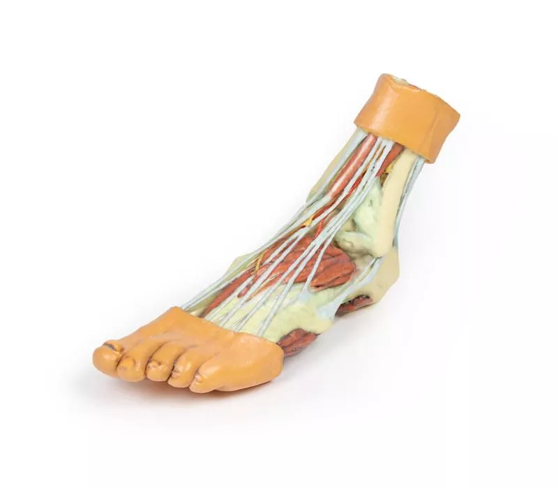
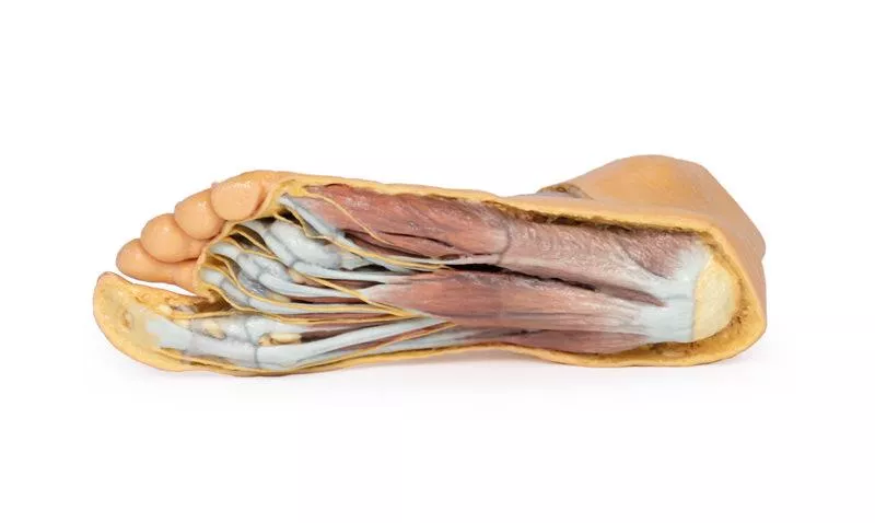
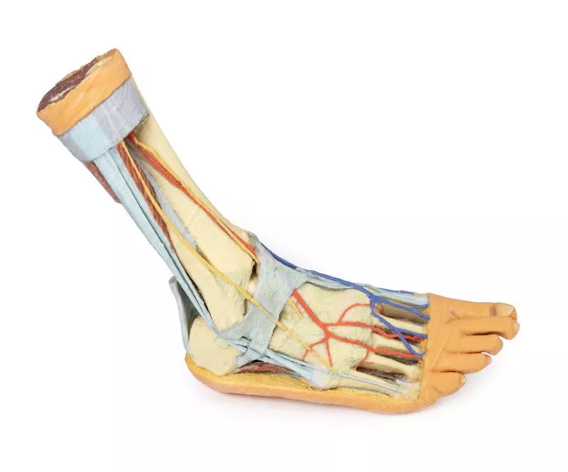
This 3D printed specimen presents a mixed superficial and deep dissection of the distal leg and foot, providing a detailed view of tendons, muscles, and neurovascular structures.
Posterior and Medial Anatomy
Posteriorly, the compartment muscles and neurovascular structures have been removed to highlight the tendocalcaneus and calcaneus body. Medially, the tibialis posterior, flexor digitorum longus, and flexor hallucis longus tendons are visible deep to the crural fascia, passing under the opened flexor retinaculum toward the medial foot. The adductor hallucis, medial head of the flexor hallucis brevis, and flexor digitorum brevis muscles are fully exposed on the medial aspect.
Dorsal Anatomy
On the dorsum of the foot, both superior and inferior extensor retinacula are preserved, with anterior compartment muscles extending to their distal attachments, including the fibularis tertius. The anterior tibial artery is visible continuing as the dorsalis pedis artery. Deep to the long tendons, the extensor hallucis brevis, extensor digitorum brevis, and dorsal interosseous muscles are clearly visible.
Lateral Anatomy
Laterally, the fibularis longus and brevis muscles are visible beneath the crural fascia, with tendons passing under both superior and inferior fibular retinacula. The abductor digiti minimi muscle is exposed along the lateral margin of the foot.
Erler-Zimmer
Erler-Zimmer GmbH & Co.KG
Hauptstrasse 27
77886 Lauf
Germany
info@erler-zimmer.de
Achtung! Medizinisches Ausbildungsmaterial, kein Spielzeug. Nicht geeignet für Personen unter 14 Jahren.
Attention! Medical training material, not a toy. Not suitable for persons under 14 years of age.


